Survivalist Pro
Photo by Mikhail Nilov Pexels Logo Photo: Mikhail Nilov

Which victim has given CPR immediately?

CPR should be performed immediately on any person who has become unconscious and is found to be pulseless.

How can I detox my body in 3 days?

A 3-day cleansing diet is a short-term diet plan that focuses on consuming whole and natural foods to help detoxify the body. It typically includes...

Read More »
How long did people live in 100 AD?

Their ages of death were compared to men listed in the more recent Chambers Biographical Dictionary. Of 397 ancients in total, 99 died violently by...

Read More »

AHA guidelines on initiating CPR

As with the adult BLS recommendations, the AHA 2010 guidelines revised the initial CPR sequence of steps from ABC (airway, breathing, chest compressions) to CAB (chest compressions, airway, breathing). [50] This change was reaffirmed in the 2020 update, which states "It may be reasonable to initiate CPR with compressions-airway-breathing over airway breathing-compressions." [43] The AHA 2020 guidelines also recommend that (1) lay rescuers should begin CPR for any victim who is unresponsive, not breathing normally, and does not have signs of life; do not check for a pulse and (2) in infants and children with no signs of life, it is reasonable for healthcare providers to check for a pulse for up to 10 seconds and begin compressions unless a definite pulse is felt. [43]

AHA guidelines algorithm for pediatric BLS

For lay rescuers

Step 1. Make sure the scene is safe. Check to see if the person is awake and breathing normally.

Step 2. If not, shout for help.

If you are alone and have a cell phone, call 911 then perform CPR (30 compressions:2 breaths) for 5 cycles (~2 minutes), then get an AED. If you are alone and do not have a cell phone, perform CPR (30 compressions:2 breaths) for 5 cycles (~2 minutes), then get an AED. If two or more people are available to help, one person calls 911 and then gets an AED, while the other person performs CPR (30 compressions:2 breaths). Step 3. Repeat cycles of CPR (30 compressions:2 breaths); use AED as soon as it arrives.

For single healthcare providers on scene

Step 1. Make sure the scene is safe. Check to see if the person is awake and breathing normally.

Step 2. If not, shout for help. Activate 911.

Step 3. Look for no breathing or only gasping and (simultaneously) check for a DEFINITE pulse WITHIN 10 SECONDS.

3b. If pulse and normal breathing, monitor until EMS arrives.

3c. If pulse but NO normal breathing:

Provide rescue breathing, at 1 breath every 2-3 seconds, or about 20-30 breaths/min. Assess pulse rate for no more than 10 seconds. If the heartrate is less than 60 beats/min with signs of poor perfusion, begin CPR. Otherwise, continue rescue breathing at 1 breath every 2-3 seconds, or about 20-30 breaths/min. Recheck the pulse every 2 minutes. 3d. If no pulse or normal breathing AND a witnessed sudden collapse, call 911, then go get an AED, then use the AED and perform CPR (30 compressions:2 breaths).

3e. If no pulse and NOT witnessed sudden collapse:

If you are alone: perform CPR (30 compressions:2 breaths) for 5 cycles (~2 minutes), then call 911 and go get an AED. If two or more people are available to help, one person calls 911 and then gets an AED, while the other person performs CPR (30 compressions:2 breaths). Step 4. Use AED as soon as it is available. If shock is advised, give 1 shock. Resume CPR immediately for 2 minutes (until prompted by AED to allow rhythm check). Continue until ALS providers take over or the person starts to move.

For two or more healthcare providers on scene

Step 1. Make sure the scene is safe. Check to see if the person is awake and breathing normally.

Step 2. If not, shout for help.

Step 3. One person calls 911 and then gets an AED, while the other person looks for no breathing or only gasping and (simultaneously) checks for a DEFINITE pulse WITHIN 10 SECONDS.

3b. If pulse and normal breathing, monitor until EMS arrives.

3c. If pulse but NO normal breathing:

Provide rescue breathing, at 1 breath every 2-3 seconds, or about 20-30 breaths/min. Assess pulse rate for no more than 10 seconds. If the heartrate is less than 60 beats/min with signs of poor perfusion, begin CPR. Otherwise, continue rescue breathing at 1 breath every 2-3 seconds, or about 20-30 breaths/min. Recheck the pulse every 2 minutes. 3d. If no pulse or normal breathing, start CPR. The first rescuer performs cycles of 30 compressions and 2 breaths. When the second rescuer returns, the two perform cycles of 15 compressions and 2 breaths. Use the AED as soon as it is available.

Step 4. Use AED as soon as it is available.

Use AED as soon as it is available. If shock is advised, give 1 shock. Resume CPR immediately for 2 minutes (until prompted by AED to allow rhythm check). Continue until ALS providers take over or the person starts to move.

AHA guidelines algorithm for pediatric ALS

Why do some people not see clutter?

One main reason is that we are so accustomed to and easily habituate to our environment, and in some ways that can help us focus. For example, you...

Read More »
What was a typical dinner in the 1930s?

A 1930s dinner party menu would probably also have included dishes that mirrored what Hollywood considered sophisticated European taste with items...

Read More »

Step 1: Begin CPR. Begin bag-mask ventilation and give oxygen. Attach monitor/defibrillator.

Step 2a: If VF/pVT, deliver shock.

Step 2b: If PEA/asystole, give epinephrine as soon as possible and go to step 8 (below). Step 3. Continue CPR for 2 min (5 rounds). Establish IV (preferred) or IO access.

Step 4a. If VF/pVT, deliver shock.

Step 4b: If PEA/asystole, give epinephrine as soon as possible and go to step 8 (below). Step 5. Continue CPR for 2 min (5 rounds). Give epinephrine every 3-5 minutes. Consider advanced airway placement.

Step 6a. If VF/pVT, deliver shock.

Step 6b: If PEA/asystole, give epinephrine as soon as possible and go to step 8 (below). Step 7. Continue CPR for 2 min (5 rounds). Continue epinephrine every 3-5 minutes. Give amiodarone (or lidocaine). Treat reversible causes. Go to step 4 (above). Step 8. Continue CPR for 2 min (5 rounds). Establish IV (preferred) or IO access. Continue epinephrine every 3-5 min. Consider advanced airway and capnography.

Step 9a. If VF/pVT, go to step 6a (above) (deliver shock).

Step 9b: If PEA/asystole, continue CPR for 2 min (5 rounds). Treat reversible causes.

Step 10a. If VF/pVT, go to step 6a (above) (deliver shock).

Step 10b: If PEA/asystole, go to step 8 (above).

If signs of ROSC are noted, go to Post–Cardiac Arrest Care

High-quality CPR

The following are considered essential elements of high-quality CPR:

Compression rate of 100-120/min

Compression depth to at least one third of the anterior-posterior diameter of the chest (approximately 4 cm in infants to 5 inches in children); for adolescents, the adult compression depth of at least 5 cm, but no more than 6 cm should be used.

Complete chest recoil after each compression

Minimized interruptions in chest compressions

Avoidance of excessive ventilation

As with BLS, algorithms are a key component of pediatric advanced life support (PALS) and are designed to simplify and expedite recognition and treatment of life-threatening conditions. Unlike BLS, PALS typically involves a coordinated team of trained responders who are able to initiate several processes simultaneously.

VF or pVT

The following summarizes the AHA PALS algorithm for VF or pVT [49] :

Call for help and activate the emergency response

Initiate high-quality CPR and give oxygen

Attach an ECG monitor and defibrillator pads

Establish vascular access; initially, attempting peripheral IV access is acceptable but only for a short, limited time; if a peripheral IV access cannot be quickly established, then an IO line should be placed by a trained provider Once the child is attached to the monitor or AED, the rhythm should be analyzed and determined to be shockable or nonshockable. Shockable rhythms include pulseless ventricular tachycardia or ventricular fibrillation. Nonshockable rhythms include pulseless electrical activity or asystole. If the rhythm indicates ventricular tachycardia or ventricular fibrillation, then it is a shockable rhythm and intervention proceeds as follows: The defibrillator should be charged to 2 J/kg, and a shock should be delivered as soon as possible once all team members are clear

Promptly restart CPR for an additional 2 minutes

Establish IV/IO access if not already done

After 2 minutes, recheck the rhythm

If the rechecked rhythm is determined to be shockable, intervention proceeds as follows: The defibrillator should be charged to 4 J/kg and a shock should be delivered

Promptly restart CPR for an additional 2 minutes

Give epinephrine 0.01 mg/kg IV or IO; this may be repeated every 3-5 minutes

Consider endotracheal intubation or other advanced airway placement

Consider amiodarone 5 mg/kg IV/IO for refractory VF/pVT (may repeat up to 2 times)

If the rhythm is nonshockable, intervention proceeds as follows:

Continue CPR for an additional 2 minutes

Establish IV/IO access

Give epinephrine 0.01 mg/kg IV/IO; this may be repeated every 3-5 minutes

Consider endotracheal intubation or other advanced airway placement

Once the patient is intubated, chest compressions and ventilations should work independently, with the compressions at a continuous rate of 100/min and the ventilations 10/min.

In addition, identify and correct the following if necessary:

Hypovolemia

Hypoxia

Hydrogen ion (acidosis)

Hyperkalemia/hypokalemia and metabolic disorders

Hypoglycemia

Hypothermia

Toxins

Tamponade, cardiac

Tension pneumothorax

Thrombosis, coronary or pulmonary

Bradyarrhythmias

Emergency treatment of bradycardia is indicated when the rhythm results in hemodynamic compromise. The AHA algorithm for the recognition and management of bradyarrhythmias is summarized below. [49] When a pediatric patient is found to be bradycardiac, quickly check for a pulse. If no pulse is found, proceed to the pulseless arrest algorithm. If a pulse is found, assess for signs of cardiopulmonary compromise. These signs include the following:

Hypotension

Can you claim an undiscovered island?

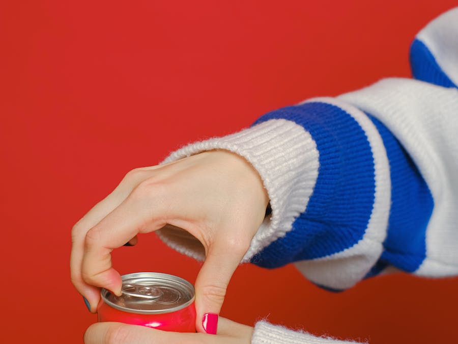
The answer is yes, but not without some difficulties. Firstly, unclaimed islands are usually unclaimed for a reason. The US retains 12 islands...

Read More »
What food is best for Bunker?

No wonder it's often called the ultimate survival food. Rice. ... Dried Beans. ... Spices. ... White vinegar. ... Canned Foods. ... Bouillon Cubes....

Read More »

Acutely altered mental status

Other signs of shock

If cardiopulmonary compromise is evident, the following immediate steps should be taken:

Put the patient on supplemental oxygen and assist ventilations as needed

Attach cardiac monitoring, blood pressure cuff, pulse oximetry, and pacing pads

Establish vascular access (IV, or IO if necessary)

Get a 12-lead ECG for rhythm analysis if possible

If the heart rate continues to be below 60 bpm and cardiopulmonary compromise is evident despite oxygenation and ventilation, then chest compressions should be initiated. While the algorithm is being applied, attempt to identify and treat any underlying causes. If bradycardia persists after 2 minutes of chest compressions, consider the following:

Epinephrine: 0.01 mg/kg IV or IO; repeat every 3-5 minutes

Atropine: 0.02 mg/kg, not to exceed 0.5 mg/dose (for increased vagal tone or primary heart block) may be repeated once

Transcutaneous or transvenous pacing

Continue to identify and treat any underlying causes

If the bradycardia resolves, continue to support the ABCs, monitor the child, and consider expert consultation. If the bradycardia evolves into pulseless arrest, proceed to the pulseless arrest algorithm.

Tachyarrhythmia

The most common types of tachycardia in the pediatric population are sinus tachycardia, supraventricular tachycardia, and ventricular tachycardia. As with other elements of PALS, an algorithmic approach is used for tachyarrhythmia, as outlined below. If a pediatric patient is found to be unresponsive and not breathing in the context of tachycardia on the monitor, then proceed to the pulseless arrest algorithm. If a pulse is found, assess for signs of cardiopulmonary compromise. These signs include the following:

Hypotension

Acutely altered mental status

Other signs of shock

If cardiopulmonary compromise is evident, the following immediate steps should be taken:

Put the patient on supplemental oxygen and assist ventilations as needed

Attach cardiac monitoring, blood pressure cuff, pulse oximetry, and defibrillator pads

Establish vascular access (IV, or IO if necessary)

Get a 12-lead ECG for rhythm analysis

Evaluate the ECG and determine if the QRS duration is narrow or wide If the QRS is wide on the initial ECG, ventricular tachycardia should be assumed. Supraventricular tachycardia with aberrant conduction is a less common possibility. If the patient shows signs of cardiopulmonary compromise, synchronized cardioversion is delivered at 0.5-1 J/kg, with an increase to 2 J/kg if initially unsuccessful. If the patient shows no signs of cardiopulmonary compromise, adenosine may be empirically given for the possibility of supraventricular tachycardia with aberrancy. Amiodarone and procainamide should not be routinely administered together, but they may be given in conjunction with expert consultation, as follows:

Amiodarone: 5 mg/kg IV infused over 20-60 minutes

Procainamide: 15 mg/kg IV infused over 30-60 minutes

If the QRS is narrow, determine whether sinus tachycardia or supraventricular tachycardia is more probable. Evidence supporting sinus tachycardia includes the following:

Presence of P waves

Variable R-R intervals

Heart rate less than 180 bpm

Evidence supporting supraventricular tachycardia includes the following:

Absence of P waves

No R-R variability

Heart rate 180 bpm or greater

Sinus tachycardia

Treat the underlying cause(s). Common causes of sinus tachycardia include hypovolemia, sepsis, fever, pain, hypoxia, and anemia. The history and physical examination can provide important information for narrowing the differential diagnosis.

Supraventricular tachycardia

While preparations are being made for chemical or electrical cardioversion, vagal maneuvers may be attempted to break the dysrhythmia. Vagal maneuvers include the following:

Application of an ice bag to the child's face

Unilateral carotid massage in older children.

If vagal maneuvers are unsuccessful and the patient has IV or IO access, then chemical cardioversion with adenosine is indicated. The regimen is as follows:

Push adenosine 0.1 mg/kg (not to exceed 6 mg)

If unsuccessful, second dose of 0.2 mg/kg (not to exceed 12 mg)

If chemical cardioversion is unsuccessful or not available, electrical cardioversion is indicated. The regimen is as follows:

If possible, sedate the patient beforehand, but do not delay cardioversion

Deliver a synchronized shock at 0.5-1 J/kg

If this is not successful, increase the charge to 2 J/kg

If chemical and electrical cardioversion continue to be unsuccessful, consider expert consultation for additional antiarrhythmics and rate-controlling recommendations.

What food can you bring on Alone?

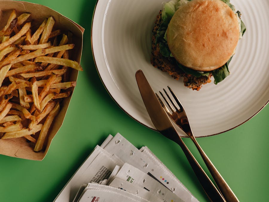
Survival tools on the list include a hatchet, climbing rope, a hammock, fishing line and a pocket knife. Contestants can even bring a couple items...

Read More »
What are the 5 signs of emotional suffering?

Common warning signs of emotional distress include: Eating or sleeping too much or too little. Pulling away from people and things. Having low or...

Read More »
What is an off the grid person?

not using public supplies of utilities such as electricity, water, etc. Oct 21, 2009

Read More »
Is it OK to keep a gun safe in the garage?

It's best to avoid placing our gun safes in the garage, but if there is no other choice, it's a good practice to bolt it to the floor and build a...

Read More »